作者: admin

  • What diseases do you see in the B-ultrasound test?

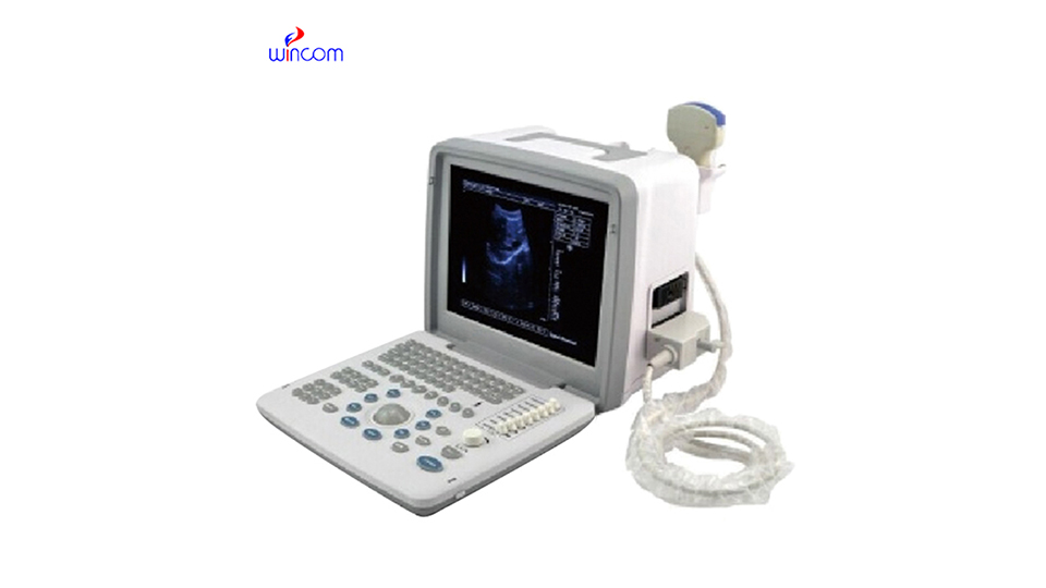
    B-ultrasound can detect liver diseases, gallbladder diseases, kidney diseases, obstetrics and gynecology diseases, and cardiovascular diseases. The specific analysis is as follows:

    1. Liver diseases: B-ultrasound can clearly display the size, shape, internal structure, etc. of the liver. For benign lesions such as liver cysts and hepatic hemangioma, a mass with clear boundaries and uniform or uneven echoes can be found in the liver by B-ultrasound. In terms of diffuse liver lesions, such as fatty liver, enhanced liver echo, enhanced front-field echo, and attenuated back-field echo can be seen under B-ultrasound. For liver cirrhosis, B-ultrasound can show signs such as reduced liver size, uneven surface, and thickened liver parenchyma echoes.

    2. Gallbladder disease: B-ultrasound is a common method for checking gallbladder disease. For gallbladder stones, on B-ultrasound images, they appear as a strong echo light mass in the gallbladder, with acoustic shadows behind them, and can move with the change of body position. In cholecystitis, the gallbladder wall will be thickened and rough, the gallbladder may be enlarged or shrunk, and the bile sound transmission will be poor. Under B-ultrasound, gallbladder polyps were shown as isoechoic or slightly hyperechoic nodules protruding from the gallbladder wall into the gallbladder cavity. They did not move with the body position and had no sound behind them.

    3. Kidney disease: In kidney examination, B-ultrasound plays an important role. For renal cysts, B-ultrasound can show a round or elliptical anechoic area in the renal parenchyma with clear boundaries and smooth cyst walls. During hydronephrosis, the renal pelvis and calyces are dilated and liquid dark areas appear. In severe cases, the kidney volume increases. For renal tumors, B-ultrasound can detect space-occupying lesions in the kidney. Depending on the nature of the tumor, the echo manifestations also vary. For example, renal cancer often presents as hypoechoic or isoechoic masses.

    4. Obstetrics and gynecology diseases: In the field of obstetrics and gynecology, B-ultrasound is widely used. In terms of pregnancy, it is possible to determine whether you are pregnant and determine the location, size, shape, etc. of the gestational sac. In the first trimester of pregnancy, B-ultrasound can detect fetal buds and fetal heart beats. For uterine fibroids, B-ultrasound showed hypoechoic or isoechoic nodules in the myometrium with clear or unclear boundaries. Under B-ultrasound, ovarian cysts appear as circular or elliptical anechoic areas in the ovary with thin and smooth walls.

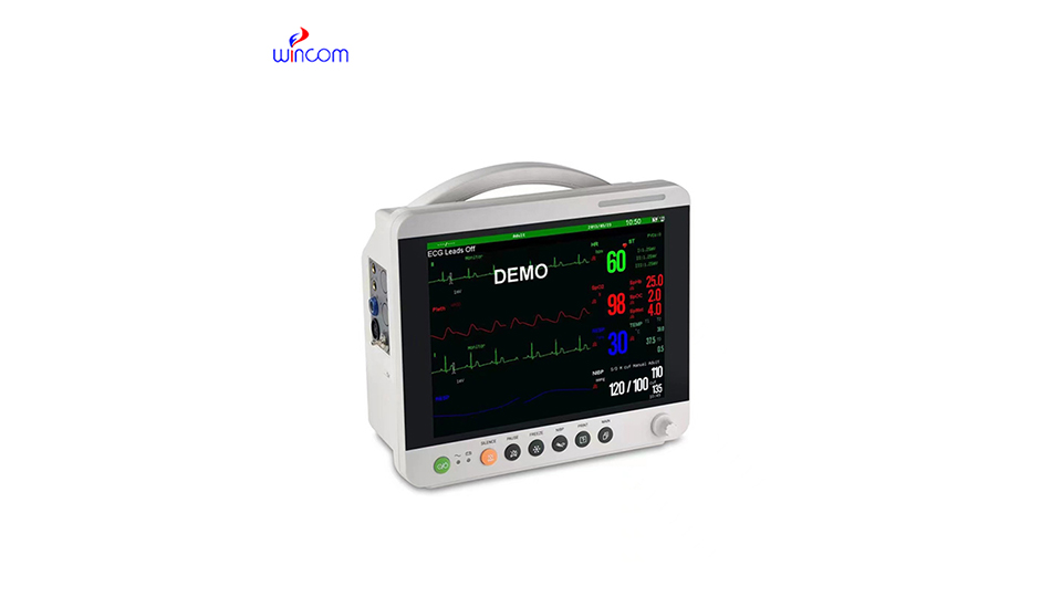
    5. Cardiovascular disease: B-ultrasound has certain value in the diagnosis of cardiovascular disease. During cardiac examination, the structure of the heart can be observed, such as the size of the atrium and ventricle, the thickness of the ventricular wall, etc. For pericardial effusion, B-ultrasound can detect the liquid dark area in the pericardial cavity, and the amount of effusion can be judged based on the depth of the dark area. In terms of blood vessels, B-ultrasound can detect the thickness of the blood vessel wall and the presence or absence of plaque formation. For example, carotid B-ultrasound can detect carotid intima thickening, plaque, etc., and can also observe blood flow and determine whether the blood vessel is narrowed.

    Keep the inspection area clean before B-ultrasound examination to avoid affecting image quality. During examination, maintain a suitable position according to the doctor’s requirements for convenience of examination. If you examine the abdomen, you generally need to be fasting, and you need to hold urine when inspecting pelvic organs such as the bladder and uterus to improve the accuracy of the examination.

  • Can I have an MRI exam during pregnancy?

    Magnetic resonance imaging (MRI) is a radiation-free imaging method, but is it safe for pregnant women?

    一. Can pregnant women have an MRI?

    1. Is MRI safe for the fetus?

    MRI does not rely on X-rays, but uses strong magnetic fields and radio waves for imaging, so in theory it will not cause radiation damage to the fetus. The American Society of Radiology (ACR) states that “non-enhanced MRI should be performed with caution during pregnancy.” However, the following principles must be followed:

    Early pregnancy (1-12 weeks): Unless in emergencies (such as suspected ectopic pregnancy, intracranial lesions), it is recommended to postpone the examination.

    Middle and late pregnancy: MRI is performed after the doctor evaluates the risks.

    2. Under what circumstances do pregnant women need an MRI?

    MRI is usually used for the following situations: First, it is used to screen for fetal abnormalities, mainly referring to brain and spinal problems that cannot be clearly diagnosed by ultrasound. Secondly, it is used to evaluate tumors or serious infections (such as pelvic abscess, etc.) in pregnant women, such as suspected stroke, placenta accreta, ruptured ectopic pregnancy, etc.

    二. Potential risks of MRI examination

    Although the MRI itself does not radiate, it should be noted that:

    1. Risks of gadolinium contrast agents (enhanced MRI): Gadolinium may enter the fetus through the placenta and is prohibited in the first trimester and used only when absolutely necessary in the second and third trimester.

    2. Potential effects of strong magnetic fields: In theory, high magnetic fields (3T MRI) may have slight thermal effects, but there is no evidence of harm to the fetus.

    3. Noise and claustrophobia: Since MRI machines are loud (similar to the sound of electric drills), it may cause anxiety for pregnant women, so it is best to wear noise-proof headphones.

    三. Preparation for pregnant women before MRI examination

    1. physician assessment

    The need for an examination must be jointly assessed by obstetricians and radiologists and signed an informed consent form to understand the possible risks.

    2. Preparation before inspection

    Wearing: Need to replace metal-free inspection clothing (to avoid affecting imaging).

    Diet: Generally, fasting is not necessary, but abdominal MRI requires fasting for 4 hours.

    Remove metal objects: jewelry, hair clips, underwear with metal buckles, etc.

    3. mentally prepared

    Pregnant women need to understand the inspection process in advance to avoid nervousness and can apply for family companionship.

    四. Matters needing attention during the inspection process

    1. Posture: In the third trimester of pregnancy, it is recommended to position on the left side to avoid supine hypotension. Use special support pads for pregnant women to improve comfort.

    2. Cooperate with breathing: Abdominal MRI may require a brief breath-holding and following the technician’s instructions.

    3. Emergency: If you feel dizzy or abdominal pain, inform your doctor immediately via the call button.

    五. Points for attention after inspection

    1. Observe fetal movement: Pay attention to fetal movement after examination, and seek medical advice promptly if there is any abnormality.

    2. Drink plenty of water (if using contrast media): Help the body expel gadolinium faster.

    3. Pending report: Results need to be interpreted by a professional doctor and follow-up plans discussed with the obstetrician.

    六. What conditions cannot an MRI be performed?

    1. Metal implants (such as cardiac pacemakers, orthopedic steel nails) are present in the body.

    2. Severe claustrophobia (consider open MRI).

    3. Unnecessary examinations in the first trimester of pregnancy (try to postpone them until after the second trimester of pregnancy).

    七. Alternative inspection plan

    If MRI risks are high, consider:

    1. Ultrasound (preferred)-radiation-free and suitable for most fetal examinations.

    2. Low-dose CT-Use only in emergency situations (such as pulmonary embolism).

    In short, MRI examinations can be performed during pregnancy, but the necessity needs to be carefully assessed, especially in the first trimester. Avoid the use of gadolinium contrast agents unless it is absolutely necessary. Before the examination, pregnant women should make psychological and physical preparations to ensure that the examination process is safe and comfortable.

  • How to view the data on the patient monitor

    Patient monitor is a type of medical equipment used to monitor heart function and electrical activity. Heart monitoring instruments can always pay attention to the patient’s heart condition, mainly including heart rate, heart rhythm, blood pressure and other data.

    1. Heart rate: refers to the number of times the heart beats per minute and is one of the most intuitive data on heart monitoring instruments. The heart rate in normal adults ranges from 60 to 100 beats per minute. If the heart rate is higher than 100 beats per minute, it is called tachycardia. Physical factors such as exercise, emotional excitement, fever, anemia, or pathological conditions such as heart disease and hyperthyroidism can all cause tachycardia. When the heart rate is lower than 60 beats per minute, it is bradycardia, which is common in athletes and sleeping, and may also be caused by diseases such as heart block.

      2. Heart rhythm: It reflects whether the rhythm of the heart’s beating is regular. Normal rhythm is sinus rhythm, which is displayed as a regular waveform on the monitor. Once a premature beat occurs, the waveform will suddenly appear in advance, and the shape is different from the normal waveform. Premature beats can be divided into atrial premature beats, ventricular premature beats, etc. Occasional premature beats may also occur in healthy people, mostly related to factors such as fatigue and alcohol consumption; however, frequent premature beats may indicate a disease in the heart. Atrial fibrillation is also a common rhythm abnormality. On the monitor, P waves disappear and are replaced by f waves of different sizes and shapes. The heart rhythm is absolutely irregular. Atrial fibrillation increases the risk of thrombosis, which can lead to serious consequences such as stroke.

      3. Blood pressure: Including systolic blood pressure (high pressure) and diastolic blood pressure (low pressure). Systolic blood pressure represents the pressure of blood on the blood vessel wall during contraction, and the normal range is generally 90- 140mmHg; diastolic blood pressure is the pressure experienced by the blood vessel wall during diastolic, and the normal range is 60- 90mmHg. Excessive blood pressure, that is, systolic blood pressure ≥140mmHg and/or diastolic blood pressure ≥90mmHg, is a manifestation of hypertension. Long-term hypertension can damage important organs such as the heart, blood vessels, and kidneys. Low blood pressure, with systolic blood pressure <90mmHg and/or diastolic blood pressure <60mmHg, may lead to dizziness, fatigue, and even shock. It is common in conditions such as blood loss, dehydration, and severe infection.

      4. Blood oxygen saturation: refers to the degree of oxygen binding to hemoglobin in the blood, which should normally be between 95% and 100%. When the blood oxygen saturation is lower than 90%, it indicates that the body is hypoxic. Lung diseases such as pneumonia, chronic obstructive pulmonary disease, and cardiac dysfunction may all cause blood oxygen saturation to drop. Continued low oxygen saturation can affect the normal function of various organs in the body, especially the brain and heart, and can be life-threatening in severe cases.

      Understanding the data on heart monitoring instruments can give you a preliminary understanding of heart health. However, it should be noted that the interpretation of data requires professional knowledge. If abnormal data is found, the doctor should be notified in time for accurate judgment and processing.

      Precautions for using patient monitor

      During the use of monitoring instruments, patients should try to avoid strenuous exercise and large limb movements to prevent electrode pads from shifting or falling off, and at the same time reduce interference with ECG signals, blood pressure measurement and other data. In addition, stay away from strong magnetic field environments, such as nuclear magnetic resonance equipment, large motors, etc., because magnetic field interference may cause abnormal fluctuations in monitoring instrument data and affect the doctor’s judgment of the condition.

      Once abnormalities are found in the monitoring instrument data, such as too fast or too slow heart rate, too high or too low blood pressure, significant drop in blood oxygen saturation, etc., immediately check whether the instrument connection is normal and whether the patient’s physical condition has changed. If the instrument failure is eliminated, the patient needs to be further examined and evaluated quickly.

    1. Common faults of X-ray machines and solutions

      As a high-precision and high-tech testing equipment, X-ray machines are widely used in medical, industrial testing, safety inspection and other fields. However, during use, the X-ray machine may encounter various faults. Below, we will introduce in detail the common faults of X-ray machines and their solutions.

      一. Common fault types and solutions

      1. Power failure: Power failure is one of the common problems of X-ray machines, manifested as failure to start normally or sudden power failure during use. For such problems, first of all, you need to check whether the power cord is connected normally and whether the power socket is firm. If the power cord is damaged, replace it promptly. In addition, the working status of the power module should also be checked regularly to ensure its stability and reliability.

      2. Image quality problems: Image blur, distortion or artifacts are image quality problems that may occur during the use of X-ray machines. Such issues are usually related to device settings, environmental factors, or hardware failures. Solutions include adjusting equipment parameters, optimizing environmental settings, and replacing damaged parts in a timely manner.

      3. Mechanical failures: Mechanical failures such as the conveyor belt stuck and poor detector movement may affect the normal operation of the X-ray machine. For such problems, the equipment should be inspected and cleaned regularly to ensure that the connections and lubrication between the various components are good. If necessary, you can ask professional personnel to repair and adjust the equipment.

      二. CL code of X-ray machine

      CL code (Classification Code) refers to the classification code of the X-ray machine and is used to indicate the type, specifications and main purpose of the equipment. Understanding CL codes helps users correctly select and use X-ray machines, and also helps maintain and manage equipment. CL codes usually consist of a string of numbers and letters, with specific meanings and rules. When purchasing and using X-ray machines, users should carefully review the equipment manual to understand the specific meaning and requirements of the CL code to ensure the correct use and safe operation of the equipment.

      三. Care and maintenance

      In order to reduce the risk of failure of the X-ray machine, users should carry out regular maintenance and maintenance work. Specific measures include but are not limited to: keeping the equipment clean and dry, regularly checking the integrity of power cords and connecting parts, timely adjusting equipment parameters to ensure image quality, and regularly replacing worn parts. In addition, users should also use equipment in strict accordance with operating procedures to avoid equipment damage or personal injury caused by improper operation.

      In short, X-ray machines may encounter various faults and problems during use, but as long as we master common fault types and solutions, and regularly maintain and maintain the equipment, we can ensure stable operation and extend service life of the equipment. At the same time, by understanding the meaning and requirements of CL codes, we can more accurately purchase and use X-ray machines that suit our needs.