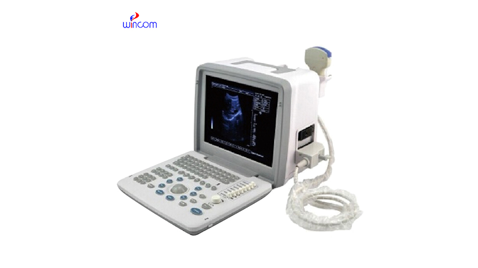
B-ultrasound can detect liver diseases, gallbladder diseases, kidney diseases, obstetrics and gynecology diseases, and cardiovascular diseases. The specific analysis is as follows:
1. Liver diseases: B-ultrasound can clearly display the size, shape, internal structure, etc. of the liver. For benign lesions such as liver cysts and hepatic hemangioma, a mass with clear boundaries and uniform or uneven echoes can be found in the liver by B-ultrasound. In terms of diffuse liver lesions, such as fatty liver, enhanced liver echo, enhanced front-field echo, and attenuated back-field echo can be seen under B-ultrasound. For liver cirrhosis, B-ultrasound can show signs such as reduced liver size, uneven surface, and thickened liver parenchyma echoes.
2. Gallbladder disease: B-ultrasound is a common method for checking gallbladder disease. For gallbladder stones, on B-ultrasound images, they appear as a strong echo light mass in the gallbladder, with acoustic shadows behind them, and can move with the change of body position. In cholecystitis, the gallbladder wall will be thickened and rough, the gallbladder may be enlarged or shrunk, and the bile sound transmission will be poor. Under B-ultrasound, gallbladder polyps were shown as isoechoic or slightly hyperechoic nodules protruding from the gallbladder wall into the gallbladder cavity. They did not move with the body position and had no sound behind them.
3. Kidney disease: In kidney examination, B-ultrasound plays an important role. For renal cysts, B-ultrasound can show a round or elliptical anechoic area in the renal parenchyma with clear boundaries and smooth cyst walls. During hydronephrosis, the renal pelvis and calyces are dilated and liquid dark areas appear. In severe cases, the kidney volume increases. For renal tumors, B-ultrasound can detect space-occupying lesions in the kidney. Depending on the nature of the tumor, the echo manifestations also vary. For example, renal cancer often presents as hypoechoic or isoechoic masses.
4. Obstetrics and gynecology diseases: In the field of obstetrics and gynecology, B-ultrasound is widely used. In terms of pregnancy, it is possible to determine whether you are pregnant and determine the location, size, shape, etc. of the gestational sac. In the first trimester of pregnancy, B-ultrasound can detect fetal buds and fetal heart beats. For uterine fibroids, B-ultrasound showed hypoechoic or isoechoic nodules in the myometrium with clear or unclear boundaries. Under B-ultrasound, ovarian cysts appear as circular or elliptical anechoic areas in the ovary with thin and smooth walls.
5. Cardiovascular disease: B-ultrasound has certain value in the diagnosis of cardiovascular disease. During cardiac examination, the structure of the heart can be observed, such as the size of the atrium and ventricle, the thickness of the ventricular wall, etc. For pericardial effusion, B-ultrasound can detect the liquid dark area in the pericardial cavity, and the amount of effusion can be judged based on the depth of the dark area. In terms of blood vessels, B-ultrasound can detect the thickness of the blood vessel wall and the presence or absence of plaque formation. For example, carotid B-ultrasound can detect carotid intima thickening, plaque, etc., and can also observe blood flow and determine whether the blood vessel is narrowed.
Keep the inspection area clean before B-ultrasound examination to avoid affecting image quality. During examination, maintain a suitable position according to the doctor’s requirements for convenience of examination. If you examine the abdomen, you generally need to be fasting, and you need to hold urine when inspecting pelvic organs such as the bladder and uterus to improve the accuracy of the examination.

